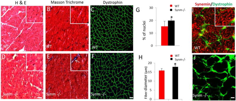
Fig. 5.
Histological changes in cardiac muscle of synm −/− and WT mice. The images show the myoplasm of cardiocytes stained with hematoxylin and eosin in pink and nuclei in blue (A,D). Masson trichrome staining revealed small regions of fibrosis (white arrow) in synm −/− heart (inset in E) (B,E). Dystrophin labeling is similar in synm −/− and controls, but the sizes of myocytes appear larger in the synm-null (C,F). Graphs show a higher number of nuclei and enlargement of cardiomyocytes in synm −/− heart (G,H). Specificity of the antisynemin antibody in cross sections of WT and synm −/− heart (I,J). *p < 0.05. Scale bars: C, F´= 20 µm; I,J= 5 µm. Insets are enlarged 2.5×. (For interpretation of the references to colour in this figure legend, the reader is referred to the web version of this article.)

