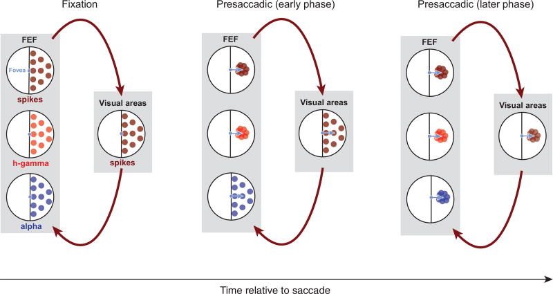
Figure 7. Possible Basis for Separable Dynamics of Spatial Information during Eye Movements.
Black circles represent the visual field; colored dots denote RFs. Dark red dots indicate spiking RFs in the recorded visual field. Red dots indicate high-gamma RFs and blue dots indicate alpha RFs. The schematic depicts that high-gamma RFs may be largely generated locally within the FEF, whereas the alpha RFs may largely reflect the synaptic input from upstream structures. Arrows indicate the signal flow between areas. Shortly before a saccade, the convergence of RFs toward the movement goal could originate first from FEF neurons and be associated with a similar convergence in the high-gamma activity. At the same time, synaptic inputs from distal sources, reflected in the alpha band, might largely maintain their retinocentric representation. Subsequently, as a result of recurrence with the FEF, visual inputs to the FEF could gradually begin to exhibit RF convergence as well.

