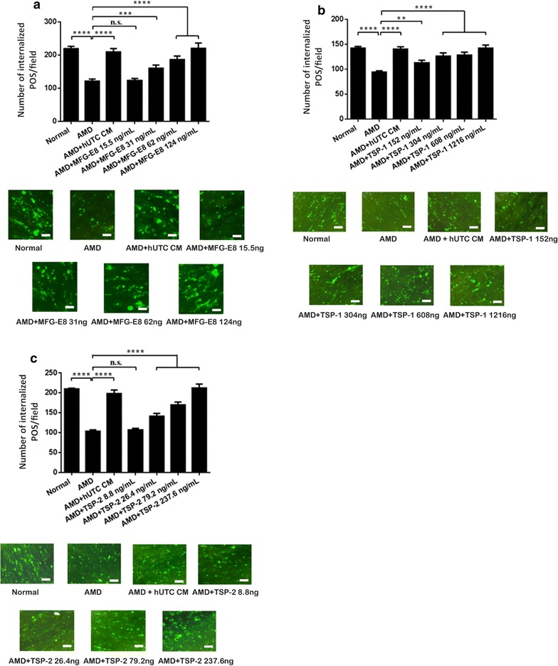
Fig. 5.

Effect of bridge molecules on phagocytosis in human RPE from donor eyes with AMD. The human RPE cells isolated from eyes of 4 AMD donors with age 65, 84, 86, and 88 were cultured as described in “Methods”. The photoreceptor OS were incubated with recombinant human MFG-E8 (a), TSP-1 (b), or TSP-2 (c) for 24 h and then fed to the RPE cells for phagocytosis assay in the absence of the hUTC CM. The POS pre-incubated with the hUTC CM was used as a positive control for the assay. Normal RPE cells from eyes of donors without ocular diseases, AMD RPE cells from eyes of donors with AMD, CM conditioned medium. Data represent the mean ± SEM (n = 4). ****P < 0.0001, ***P < 0.001, **P < 0.01, ns not significant. Representative images of RPE containing fluorescent ingested POS are shown (scale bar = 10 µm)
