Figure 2.
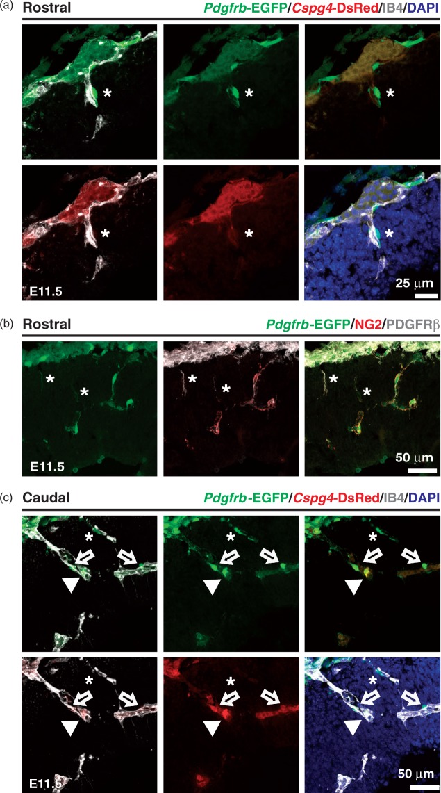
Pdgfrb-EGFP-, and Cspg4-DsRed expression in cerebral cortex vasculature. Rostral (a and b) and caudal (c) regions of the forebrain cortex from Pdgfrb-EGFP; Cspg4-DsRed (a and c) and Pdgfrb-EGFP (b) mice at E11.5. Pdgfrb-EGFP (b) mouse brain sections were stained with PDGFRβ (white) and NG2 (red). A vascular mural cell (Pdgfrb-EGFP+ /Cspg4-DsRed−) detected in vicinity to the IB4+ vessel (asterisks, a and c), cellular processes displaying Pdgfrb-EGFP with PDGFRβ+ and NG2- immunoactivity (asterisks, b), a cell showing strong Pdgfrb-EGFP and weak Cspg4-DsRed signals (open arrows, c), and a cell with both Pdgfrb-EGFP+ and Cspg4-DsRed+ signals (arrowheads, c) are shown.

