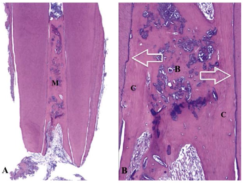
Figure 2.
Histology of revascularized human immature permanent tooth (hematoxylin-eosin stain). (A) The canal space filled with mineralized tissue (M) (original magnification ×16); (B) High magnification of A. The mineralized tissue similar to bone (B) and cementum (C). The canal dentin walls covered by newly formed cellular cementum-like tissue (arrows) (original magnification ×100) [53].

