
Figure 3. Fibrinogen Disrupts OPC Differentiation through BMP Ligand-Independent Activation of ACVR1.
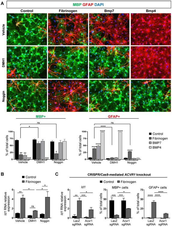
(A) Immunofluorescence for MBP (green) and GFAP (red) in primary rat OPCs treated with fibrinogen, BMP7, or BMP4, and DMH1, noggin, or vehicle control. Nuclei are stained with DAPI. Data are mean ± s.e.m. from n = 2-3 independent experiments. ns = not significant, *p < 0.05, **p < 0.01, ***p<0.001, ****p < 0.0001 (two-way ANOVA with Bonferroni). Scale bar: 50 μm.
(B) Id1 in primary rat OPCs treated with fibrinogen and DMH1, noggin, or vehicle control. Values are mean ± s.e.m. from n = 4–7 wells from 2-3 independent experiments. ns = not significant, *p < 0.05, **p < 0.01 (two-way ANOVA with Bonferroni).
(C) Analysis of primary rat OPCs transfected with a Cas9 expression plasmid containing single-guide RNA (sgRNA) for either LacZ (control) or Acvr1. Left: Id1 after 2h fibrinogen treatment, n = 3 independent experiments. Right: Quantification of MBP+ and GFAP+ cells after 3 day fibrinogen treatment, n = 4 wells from 2 independent experiments. Values are mean ± s.e.m. *p < 0.05, **p < 0.01, ***p<0.001, ****p < 0.0001 (two-way ANOVA with Holm-Sidak).