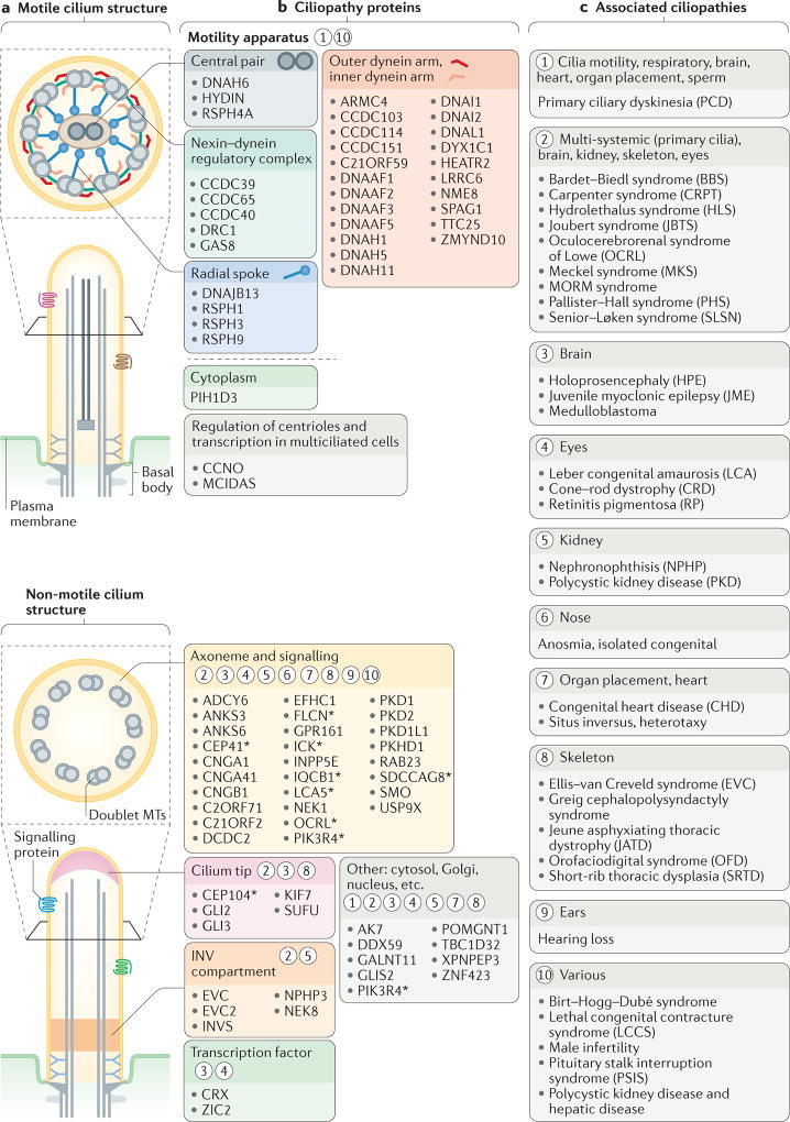
Figure 3. Structural and functional features of motile and sensory cilia are associated with ciliopathies.
a | The major structures of motile and non-motile cilia (also see FIG. 1). b | Major sites of action for ciliopathy-associated proteins that are components of motile cilia (motility apparatus or transcription factors required for the generation of motile cilia) and sensory cilia (axonemal and signalling proteins, ciliary tip proteins or inversin (INV) compartment proteins). The asterisks indicate proteins that are also localized to other ciliary regions during ciliogenesis (shown in FIG. 4) or ciliary trafficking (shown in FIG. 5). Circled numbers indicate one or more ciliopathies that result from defects in the different ciliary compartments and proteins. c | Ciliopathies grouped into major categories that are associated with the proteins and ciliary regions shown in part b.

