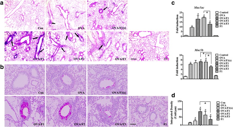
Fig. 6.

Exposure of infant mice to PM enhances pulmonary inflammation and airway mucus secretion in an asthma model. a Morphologic features of mouse lung inflammation stained with H&E. Arrows depict peribronchiolar inflammation. Scale bar = 50 μm. b Representative micrographs of PAS-stained mucus in airway epithelium. Scale bar = 100 μm. c The mRNA expression of Muc5ac and Muc5b in mouse lung tissues. d The degree of PAS staining in the airway epithelium was quantified using ImagePro-Plus 7.0 (Media Cybernetics, USA). Data are plotted as means ± SEM (n = 5 mice/group). *p < 0.05 vs. Con group; #p < 0.05 vs. OVA group, &p < 0.05 vs. OVA/PM (F1, F2 or F3)
