. 2017 Jun 13;8(8):5335–5344. doi: 10.1039/c7sc01932g

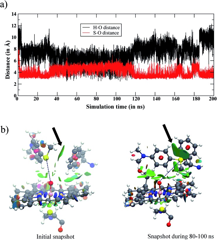
Fig. 2. (a) The variation of the Fe Created by potrace 1.16, written by Peter Selinger 2001-2019 O···H and Fe Created by potrace 1.16, written by Peter Selinger 2001-2019 O···S distances vs. simulation time for the WT enzyme. Initially, the amine and sulfur ends of Sub1 are far away from iron-oxo, however, in the later stages both ends become accessible to Cpd I. However, the major basin of the snapshots places the sulfur end closest to Cpd I. (b) The structures in the initial snapshot and the one during the 80–100 ns of the MD trajectory are shown along with the noncovalent interactions of Sub1 with Phe87 and Cpd I. The green surfaces show the favorable noncovalent interactions and the area of the green patches indicates the interactions’ intensity. Note the increase in the intensity between the sulfur atom of Sub1 and Phe87 (highlighted by arrows) from the initial trajectory to the one in the 80–100 ns interval.

Fig. 2