
Fig. 2. Ex vivo collagen-fibrin gel cultures maintains the immune suppression of in vivo tumor microenvironment.
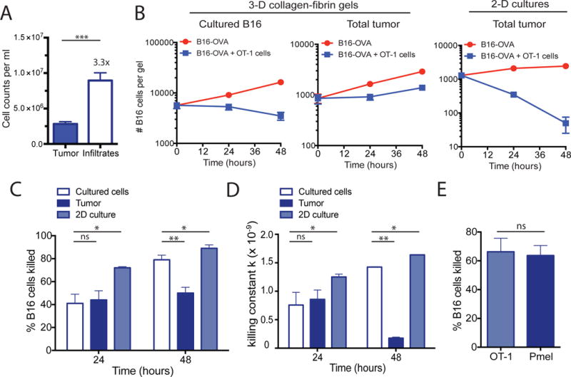
(A to E) B16-OVA tumors were excised and digested with collagenase and then disaggregated mechanically into single cell suspensions. Dissociated tumors were co-embedded in collagen-fibrin gels with in vitro–activated OT-1 cells at a 50:1 effector to target ratio. (A) The numbers of viable tumor cells and immune cell infiltrates isolated from dissociated 10-day B16-OVA tumors were determined. Data are means ± SEM of 8 experiments. (B) The numbers of viable melanoma cells recovered from the gels at the indicated times were measured. Data are means ± SEM of 3 independent experiments performed in duplicate. (C) The percentages of B16 cells killed were determined. Data are means ± SEM of 8 experiments as performed in (A). (D) The calculated value of k ± SEM from the experiments performed in (A) using the equation bt=b0 e−kpt+gt as described in Materials and Methods. (E) The percentages of B16 tumor cells killed were determined. Data are means ± SEM at 24 hours using equivalent numbers (5 × 10⁵ cells/gel) of OT-1 or Pmel CD8+ T cells in collagen-fibrin gel co-cultures of B16-OVA cells. *P ≤0.05 and **P ≤0.01. ***P ≤ 0.005.