
Fig. 4. TGF-β blockade reverses the suppression of tumor cell killing ex vivo.
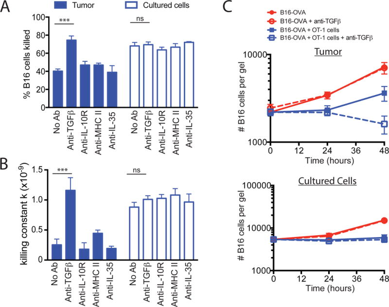
(A to C) B16-OVA tumors were excised and dissociated as described in Fig. 2. The dissociated tumors were co-embedded in collagen-fibrin gels with in vitro–activated OT-1 cells at a 50:1 effector to target ratio in the presence or absence of blocking antibodies against TGF-β, the IL-10R, MHC class II, or IL-35 (all at 10 μg/ml). (A) The percentages of B16 cells killed at 48h were determined. Data are means ± SEM of 3 experiments performed in duplicate. (B) The mean values of k ± SEM from the experiments performed in (A) were determined. (C) The mean number of clonogenic B16 remaining at the indicated times were determined. Data are means ± SEM of 3 experiments performed in duplicate. ***P ≤0.005.