
Fig. 6. Adding back Tregs to depleted tumors partially restored the suppression.
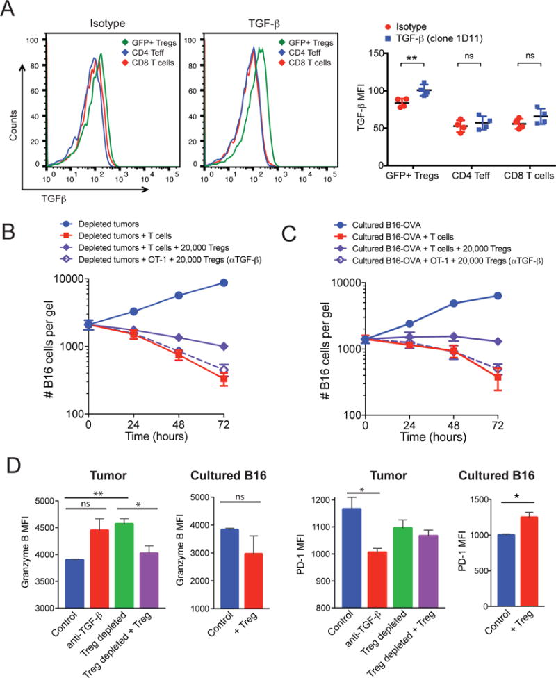
(A) B16-OVA tumors from non-depleted mice were excised and dissociated as described in Materials and Methods. Left: The cell surface expression of TGF-β on CD4+ effectors, CD8+ T cells, and CD4+Foxp3+ (GFP+) Tregs was assessed by flow cytometry. Representative histograms for the isotype control and anti-TGF-β antibody are shown. Right: Data represent the mean fluorescence (MFI) ± SEM for TGF-β relative to that or an isotype control antibody for four mice per group. (B and C) GFP+ Tregs from tumors in Foxp3-GFP mice were sorted by FACS from non-depleted tumors and pre-incubated with blocking antibody against TGF-β. Tregs (2 × 10⁴ cells) were then co-embedded with dissociated Treg-depleted B16-OVA tumors (B) or cultured B16-OVA cells (C). At the indicated times, the gels were dissolved and the numbers of remaining B16 cells were measured using a clonogenic assay. Data are means ± SEM of the numbers of B16 cells from 3 experiments performed in duplicate. (D) OT-1 CD8+ T cells were recovered from collagen-fibrin gels at 48 hours after culture under the indicated conditions and were analyzed by flow cytometry to determine the relative abundances of granzyme B (left) and PD-1 (right). Data are means ± SEM of the MFIs from triplicate analyses. *P ≤0.05 and **P ≤0.01.