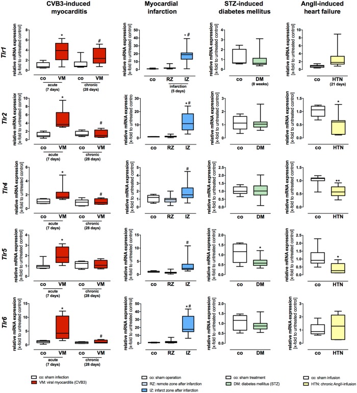
Fig 2. Gene expression levels of the plasma membrane localized TLRs Tlr1, Tlr2, Tlr4, Tlr5, and Tlr6 in different heart failure models of diverse etiology.
Cardiac tissue of healthy and diseased C57BL/6J mice was used for TaqMan based gene expression analysis of the plasma membrane localized TLRs Tlr1, Tlr2, Tlr4, Tlr5, and Tlr6. Expression levels of cardiac tissue from control mice are shown as white boxes, from diseased animals as red, blue, green or yellow boxes corresponding to the analyzed heart failure model. In viral-induced myocarditis (shown in red), gene expression of Tlr1, Tlr2, Tlr4, Tlr5, and Tlr6 was highly increased 7 days after infection compared to healthy controls. However, Tlr2 and Tlr6 displayed the highest increase during acute myocarditis. The initially increased gene expression levels returned almost with exception to basal levels 28 days after infection. In the model of myocardial infarction (shown in blue), gene expression of Tlr1, Tlr2, Tlr4, Tlr5, and Tlr6 was highly increased 5 days post infarction in the infarction zone when compared to the remote zone. In the remote zone, no increased TLR gene expression was observed. In STZ-induced diabetic cardiomyopathy (shown green), a decreased gene expression of Tlr5 in comparison to their healthy controls was detected; the remaining plasma membrane localized TLRs displayed no changes in gene expression levels. In the heart failure model caused by chronic AngII-infusion for 21 days (shown in yellow), all plasma membrane localized TLRs displayed a significant decrease when compared to their controls. Data are presented in box plots as relative mRNA expression in fold change to the corresponding untreated control using the formula 2−ΔΔCt. * = significantly different compared to corresponding control; # = significantly different compared to VM (acute—7 days) or RZ (remote zone).

