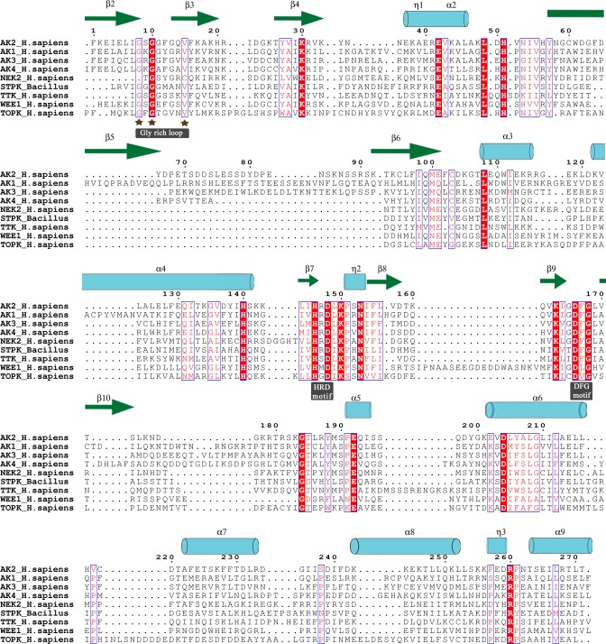
Fig 3. MSA of EIF2AKs.
Conservation plot of EIF2AK alignment is shown in alignment plot with out-group sequences of NEK2, TTK, WEE1, TOPK and STPK. Identical residues are highlighted in red background and similar residues are colored red. PDB structure 2A1A of human EIF2AK2 kinase domain is used as structural reference. The α-helices, β-strands and 310-helix are rendered with arrows, medium and small squiggles and numbered with the symbols α, β and η symbols.

