. 2018 Mar 7;7:e32723. doi: 10.7554/eLife.32723

Figure 4. Lateral interactions in TANGO1 ring assembly mediated by TANGO1 self-association.

(A, B) Co-immunoprecipitation of TANGO1-FLAG with the indicated constructs in HEK293T cells. Lysates and immunoprecipitated samples were probed for HA, FLAG and cTAGE5. 2H5 cells co-transfected with collagen VII and (C) TANGO1ΔCC1 (16 cells imaged), (D) TANGO1Δ1255–1295 (15 cells imaged) or (E) TANGO1Δ1296–1336 (16 cells imaged), were imaged by STED microscopy. (F) Schematic of interactions between TANGO1, TANGO1-short and cTAGE5. Scale bars (C–E) 2 μm; insets 200 nm.

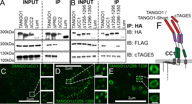
Figure 4.

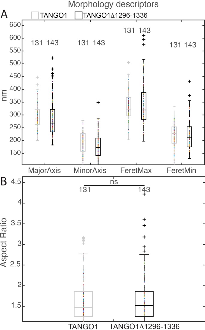
Figure 4—figure supplement 1. Morphological quantification of structures formed by TANGO1Δ1296–1336.

Figure 4—figure supplement 1.

(A) Quantification of size of TANGO1 and TANGO1Δ1296–1336, shown as a scatter plot and box plot of measured morphological descriptors: major axis and minor axis of a fitted ellipse, maximum and minimum Feret’s diameters. (B) Quantification of shape, shown as a scatter and box plot of the aspect ratio between the major and minor axes. 131 and 143 rings respectively were analysed for the two constructs. ns not significant.