
Figure 7. Chemical Inhibition of GSK3α Promotes Neural Differentiation of ESCs.
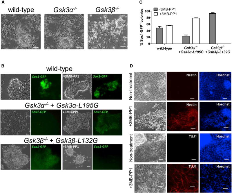
(A) Representative images of the indicated ESCs cultured in PD03/N2B27 for 9 days. Scale bars, 100 μm.
(B) Representative phase-contrast and fluorescent images of the indicated ESCs cultured in N2B27 medium only (the leftmost two columns) or N2B27 medium supplemented with 3 μM 3MB-PP1 (the rightmost two columns). ESCs were plated at clonal density and cultured in LIF/serum for 1 day. The medium was then changed to either N2B27 or 3MB-PP1/N2B27 and cultured for another 6 days before the images were taken. Scale bar, 100 μm.
(C) Quantification of Sox1-GFP positive colonies from experiments described in (B). Data represent means ± SD of three biological replicates.
(D) Representative phase-contrast and immunofluorescence staining images of Gsk3α−/− + Gsk3α-L195G 46C ESCs cultured in N2B27 medium with or without 3 μM 3MB-PP1 for 11 days. Scale bar, 100 μm.