Figure 1.

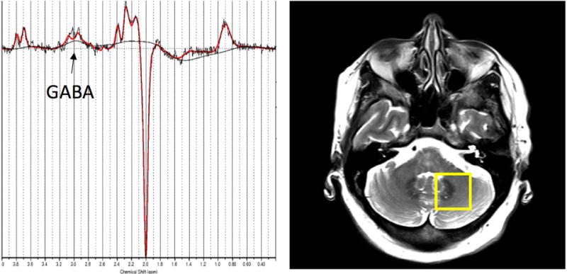
Left: Representative GABA-edited spectrum from the dentate VOI showing the raw data (black) and the LCModel fit (red). Right: Placement of the GABA VOI, containing the left cerebellar dentate.

Left: Representative GABA-edited spectrum from the dentate VOI showing the raw data (black) and the LCModel fit (red). Right: Placement of the GABA VOI, containing the left cerebellar dentate.