
Figure 2. BKCa channel expression in UAs.
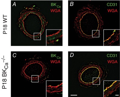
Immunohistochemistry showing expression of BKCa channel (green) in non‐pressurized P18 UAs from WT (A) and BKCa −/− (C) mice. CD31 (green) was used as an endothelial layer marker (B and D). Wheat germ agglutinin (WGA, red) was used to highlight vessel structure in all panels. Insets show high‐magnification views of boxed areas. Bars = 50 μm and 10 μm (insets).