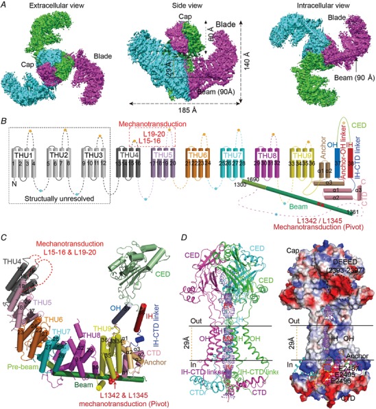
Figure 1. Structure and topology of the mechanosensitive Piezo1 channel.

A, the three‐bladed, propeller‐like cryo‐EM structure of the Piezo1 ion channel. B, nine repetitive transmembrane helical units (THUs) and the 38‐TM topology model. C, a cartoon model showing one subunit with featured structural domains labelled. D, the ion‐conducting pore module shown as a ribbon diagram (left) and surface electrostatic potential (right). The functionally identified regions and residues critical for mechanical activation of Piezo1 are indicated in B and C. Figure reproduced with permission from Zhao et al. (2018).
