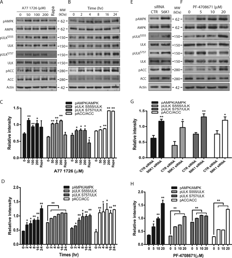
Fig. 3. AMPK and ULK1 phosphorylation by S6K1 inhibition.
NSC34 cells were incubated in complete DMEM medium in the absence or presence of the indicated concentrations of A77 1726 for 16 h (a, c) or in the presence of A77 1726 (200 μM) for the indicated time (b, d). NSC34 cells were transfected with S6K1 siRNA and incubated for 48 h (e, g) or were treated with the indicated concentrations of PF-4708671 for 16 h (f, h). Cell lysates were analyzed for total and phosphorylated proteins by western blot. The expression levels were analyzed by quantification of the density of the protein bands with NIH Image-J software and presented as bar graphs (c, d, g, h). *p < 0.05; **p < 0.01, compared to the untreated control

