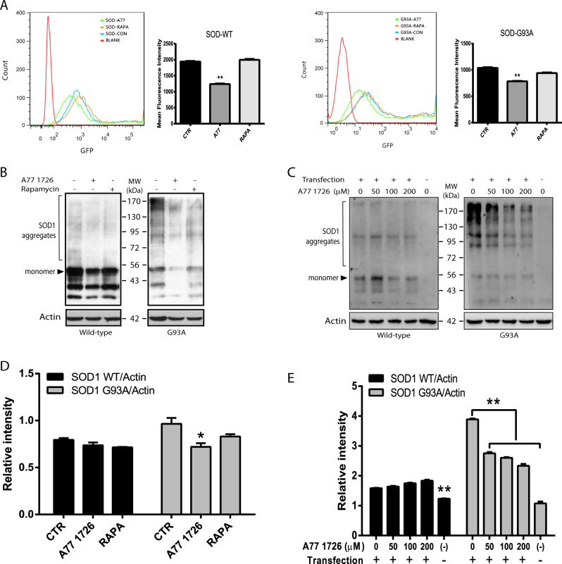
Fig. 7. Evidence that A77 1726 induces SOD1 protein degradation.
a NSC34 cells seeded in 60 mm dishes were transfected the SOD1-GFP or SOD1G93A-GFP expression vectors and treated with A77 1726 (200 μM) or rapamycin (50 nM). Single-cell suspensions were analyzed for GFP expression in a flow cytometer. The fluorescence intensity was analyzed by using FlowJo software. The results represent the mean ± SD from three independent experiments. *p < 0.05; **p < 0.01. b, c Western blot analysis of SOD1 aggregates. SOD1-GFP or SOD1G93A-GFP-transfected NSC34 cells were treated with A77 1726 or rapamycin as above (b) or treated with the indicated concentrations of A77 1726 (c) for 24 h. Insoluble fractions of the cell lysates were analyzed by western blot with an anti-SOD1 rabbit serum or actin. Protein aggregates as marked were analyzed by using a NIH Image J software and presented as bar graphs (d, e). *p < 0.05; **p < 0.01

