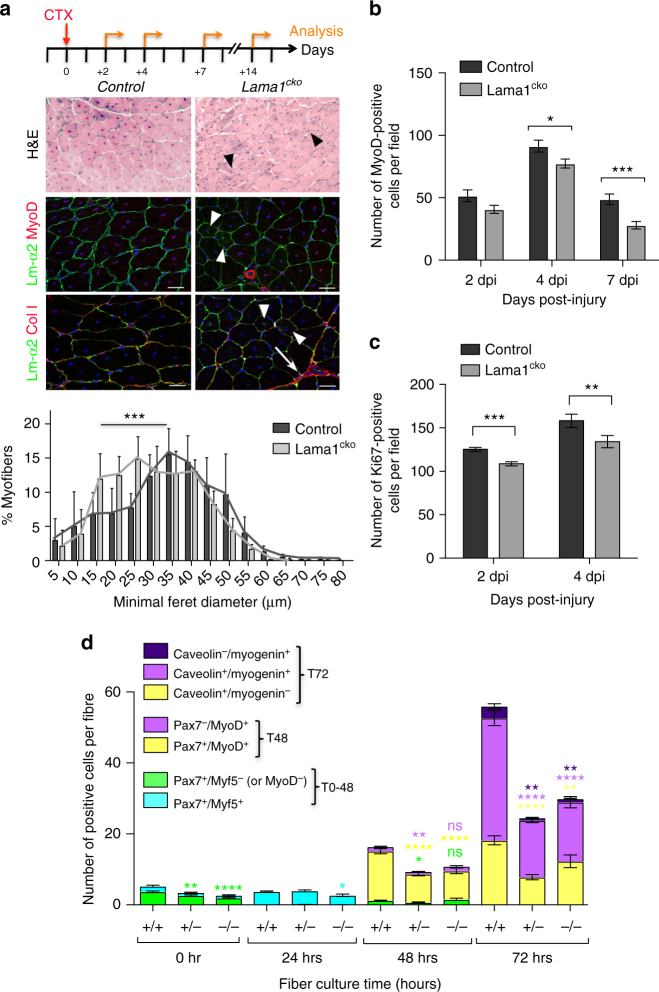
Fig. 4.
Laminin-α1 is essential for SC proliferation. a Control and Lama1cko Tibialis anterior (TA) muscle analyzed at 14 days post injury by Haematoxylin and Eosin staining, and immunofluorescence for laminin-α2 (green), MyoD (red), and collagen I (red). Black and white arrowheads indicate the presence of smaller regenerated myofibers in Lama1cko mice. White arrows indicate sites of fibrosis. The graph shows the minimal Feret diameter analysis of control and Lama1cko TA muscles at 14 dpi. Scale bar, 50 μm. n = 3 per genotype. b Number of MyoD+ cells in injured TA muscles at 2, 4, and 7 dpi in control and Lama1cko mice. n = 3 per genotype. c Number of Ki67+ cells in injured TA muscles at 2 and 4 dpi in control and Lama1cko mice. d Quantification of the number of satellite cells activated (blue), proliferating (yellow), self-renewing (green), and differentiating (purple) in a 72-hour ex-vivo culture of control, heterozygous, and Lama1cko myofibers. n = 3 per genotype with 31–79 myofibers per time point. Graphs show mean + sem. *P < 0.05, **P < 0.01, ***P < 0.001, ****P < 0.0001 (t-test for b, c, and d, and one-way ANOVA for a)

