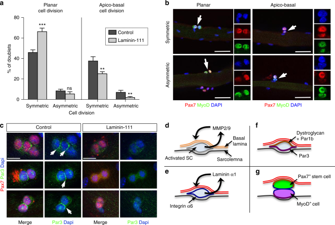
Fig. 8.
Laminin-111 treatment alters SC cell polarity and stimulates planar cell division. a Quantification of planar and apico-basal cell divisions (symmetric and asymmetric) based on the expression of Pax7 and MyoD in T46 myofibers treated with PBS (control; dark gray) or laminin-111 (light gray). n = 3 with 50–82 doublets analyzed per culture. *P < 0.05, **P < 0.01, ***P < 0.001 (t-test). b Representative immunofluorescence images of planar and apico-basal cell divisions in T46 myofibers analyzed using antibodies against Pax7 (red) and MyoD (green). White arrows indicate cell doublets. Individual color channels are shown. Scale bar: 50 μm c Representative immunofluorescence of Par3 (green) and Pax7 (red) in myofibers cultured for 46 h in in the presence of PBS (control) or laminin-111. Arrows indicate Par3 asymmetric distribution in control, but not in laminin-111 treated fibers. The white star indicates background staining. Scale bar: 10 μm. d–g Proposed model for laminin-111 control of SC self-renewal: d Upon activation, SCs upregulate MMP2 and MMP9 expression, leading to a local digestion of the laminin-α2-containing basal lamina (double orange line) at the SC niche. e Simultaneously, SCs re-express laminin-α1, which is secreted and deposited into the SC basal lamina (double red line), and the laminin-α1 receptor, integrin-α6 (blue line). f Laminin-α1-mediated signaling initiates or maintains SC polarity through the asymmetric distribution of the basal determinant Par1b (brown) and the apical determinant Par3 (purple), leading to apico-basal cell polarity. g Apico-basal asymmetric cell division yields two distinct daughter cells, including a self-renewing SC associated with the basal lamina (Pax7+ in green) and a differentiating SC (MyoD+ in purple)

