Figure 1.
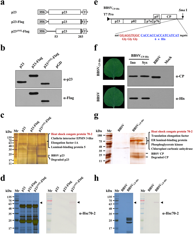
Hsc70-2 was co-purified with both BBSV p23 and CP. (a) Schematic representation of plasmids used for immunoprecipitation assays. Flag-tag (dotted rectangles) was engineered to the C-terminus of different p23 derivatives. (b) Western blot analysis of p23 and its derivatives in the infiltrated leaves of N. benthamiana. Leaves infiltrated with pGD empty vector served as the negative control. (c) Silver-stained SDS-PAGE gel image of p23 immunoprecipitates used for LC-MS/MS analysis. (d) Silver-stained SDS-PAGE and Western blot analyses of the p23 immunoprecipitates. (e) Schematic representation of the fusion of three glycine residues plus six His residues to the C-terminus of BBSV CP. (f) Analysis of the stability of His-tag during the infection of recombinant BBSV. Left panels: Symptom images of the upper uninoculated N. benthamiana leaves at 9 days after inoculation of the in vitro transcripts of BBSV and its derivative. Right panels: Western blot analysis of CP expression in the inoculated leaves (Ino) and upper uninoculated leaves (Sys) of N. benthamiana. Mock and wild-type BBSV-inoculated N. benthamiana plants served as negative and positive controls, respectively. (g) Silver-stained SDS-PAGE gel image of CP immunoprecipitates used for LC-MS/MS analysis. Note: all of the three lanes were cropped from two separate gels (see Supplementary Fig. S2). (h) Silver-stained SDS-PAGE and Western blot analyses of the CP immunoprecipitates.

