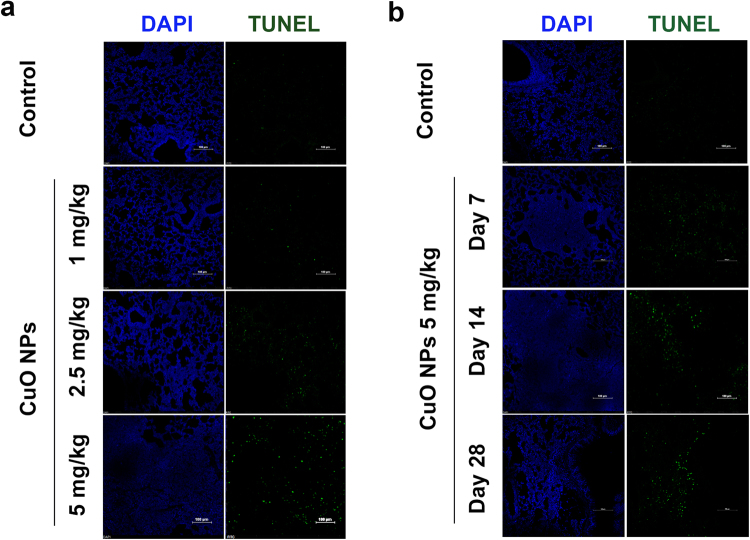
Figure 3.
Cell apoptosis in CuO NP-treated lung tissues. Cell apoptosis was determined by TUNEL staining after CuO NP treatment (n = 5 per group) compared to the saline control (n = 3). DAPI was employed for counter staining and pictures were taken using DAPI (left row), and FITC (right row) filters. The results showed that the dose-dependent manner of TUNEL staining compared to saline controls after treating the mice for 14 days (a). In the different time treatment of 5 mg/kg CuO NPs groups, the number of TUNEL positive cells reached a maximum at Day 28 compared with the other groups. Scale bar = 100 μm (b).

