Fig. 4.
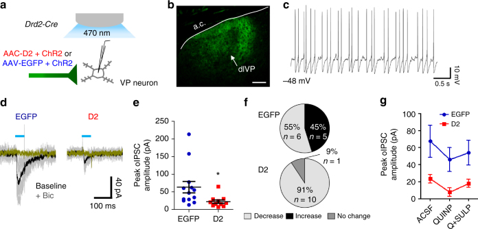
D2R upregulation reduces D2-MSN inhibitory transmission to ventral pallidum (VP). a Schematic of oIPSCs recordings from VP neurons in slice following blue-light stimulation of terminal fields from NAc MSNs co-expressing ChR2 and D2R or ChR2 and EGFP. b VP neurons were recorded from within virus-positive NAc D2-MSN terminal fields in the dorsolateral VP (dlVP). Scale bar = 150 µm. c Target neurons in VP typically displayed spontaneous firing activity, depolarized RMP, slow ramp-like depolarization preceding short duration spikes, and prominent afterhyperpolarization. d Individual (gray) and average (black) oIPSC traces (gray) from representative MSNs following five 50-ms light pulses (1 Hz). Bicuculline (10 µM) eliminated the oIPSC. e Peak oIPSC amplitude under basal conditions was significantly reduced when D2Rs were overexpressed in D2-MSNs MSNs [n = 13 cells (10 mice) or 11 cells (7 mice)]. f Quinpirole (1 µM) bidirectionally modulated oIPSCs recorded in EGFPNAcInd VP neurons MSNs [n = 11 cells (9 mice) or 10 cells (6 mice)]. In contrast, quinpirole decreased the oIPSC amplitude in >90% of cells recorded in D2R-OENAcInd VP. g Opposite influences of D2 agonist and antagonist are highlighted by the overall effect of quinpirole and quinpirole + sulpiride (1 µM) on oIPSC amplitude. Error bars = s.e.m.

