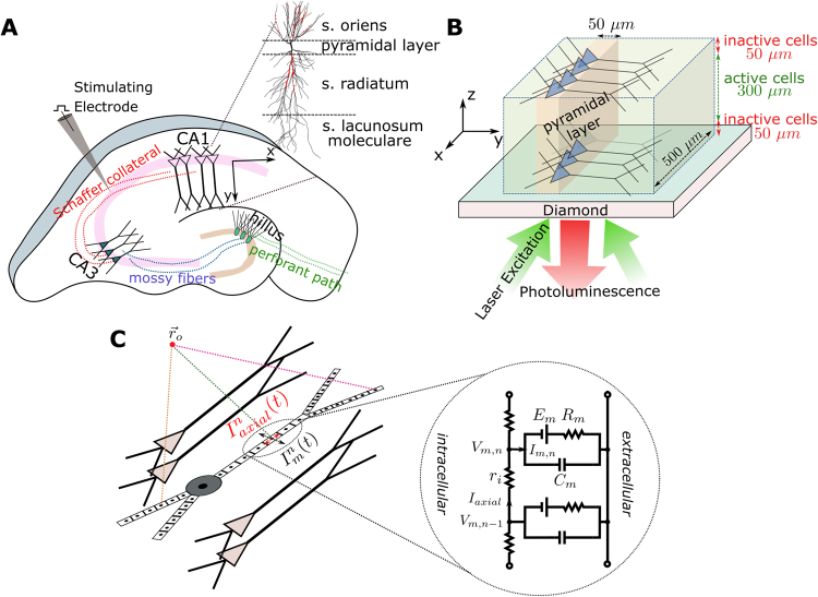
Figure 1.
(A) Illustration of a hippocampus slice with its trisynaptic path. We consider the recording of neural activity from the CA1 area that is evoked by the electrical stimulation of the Schaffer collaterals. (B) Schematic illustration of the simulated CA1 subarea with a size of 500 × 500 × 300 µm3 placed on the diamond sample. It is assumed that the neural cells in a distance of up to 50 µm to the diamond are dysfunctional due to the preparation. The pyramidal cells are equally distributed in the patch along the X and Z directions, and their soma locations are randomly jittered in a 50 µm wide band in Y direction. The changes in photoluminescence emitted by the NV-layers in the diamond and caused by the neural magnetic fields can be recorded using an arrangement similar to the inverted microscope used by Barry and colleagues10, with the camera replacing the photodetector. (C) Schematic illustration of the multi-compartment model of a pyramidal cell, as applied in our forward modeling scheme.

