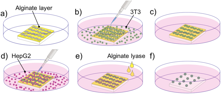
Figure 1.
Processes of seeding and culturing cells on the microplates. (a) The glass substrate with microplates was placed in a petri dish. (b) NIH/3T3 cells were seeded on the microplates, and non-adherent cells were washed away. (c) Adherent NIH/3T3 cells were cultured for 24 h. (d) HepG2 cells were then seeded onto plates and non-adherent cells were washed away. (e) The attached HepG2 cells were cultured 4 h on the NIH/3T3 cells which loaded on the microplates. (e,f) After adding alginate lyase, the microplates were folded, and a number of 3D cell co-culture microstructures were formed.

