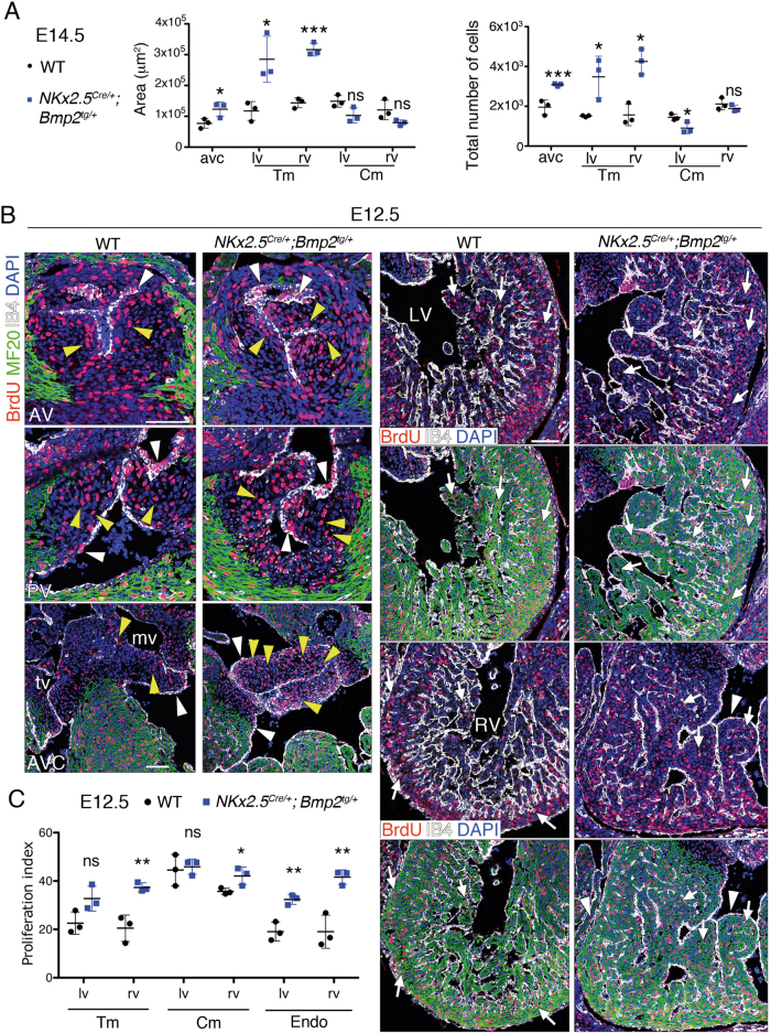
Fig. 4. Nkx2.5Cre/+;Bmp2tg/+ embryos show increased cardiac proliferation.
a Area occupied by the various cardiac territories, and the total number of cells within them, analysed in E14.5 WT and Nkx2.5Cre/+;Bmp2tg/+ hearts. b Proliferation analysis in E12.5 WT and transgenic heart sections. Sections are stained with an anti-BrdU (red) and anti-MF20 (green, myocardium) antibodies, isolectin B4 (white, endocardium) and counterstained with DAPI (blue). AV aortic valve, AVC atrioventricular valves, LV left ventricle, mv mitral valve, RV right ventricle, tv tricuspid valve. The top rows in the column corresponding to the sections of the ventricles show only anti-BrdU, IB4 and DAPI staining, to facilitate identification of BrdU-positive nuclei. The white and yellow arrowheads indicate BrdU-positive endocardium or mesenchyme nuclei, respectively. The white arrows indicate BrdU-positive nuclei in myocardium. Scale bars 200 μm. c Proliferation index in E12.5 WT and transgenic hearts is the ratio of BrdU-positive nuclei to the total number of nuclei in each cell type. t test, *P < 0.05; **P < 0.01; ns non-significant

