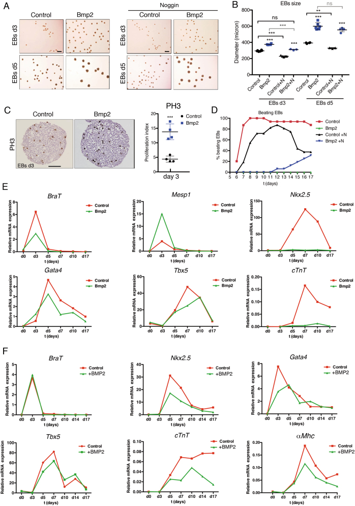
Fig. 6. Constitutive Bmp2 expression leads to increased proliferation and blockade of cardiac differentiation in embryoid bodies (EBs).
a Images showing EBs size at day 3 (d3) and day 5 (d5) of culture. Bmp2-expressing-EBs (Bmp2) are visibly larger than controls. Noggin (500 ng/ml, right panels) reduced the size of both control and Bmp2-EBs at d3 but not the size of Bmp2-EBs at d5. Scale bars 1 mm. b Size quantification of the EBs shown in a (control, WT EBs; Bmp2, Bmp2-EBs;+N, Noggin-treated EBs). Bmp2-EBs were larger than WT EBs at d3 and d5 (Bmp2-EBs vs. Control EBs). Noggin significantly reduced the size of control and Bmp2-EBs at d3 but not of d5 Bmp2-EBs at d5 (treated EBs vs untreated EBs). Noggin-treated Bmp2-EBs were similar in size to untreated WT EBs at d3 but not at d5. ***P < 0.005, **P < 0.01, ns, non-significant one-way ANOVA (non-parametric) and Bonferroni post-test. c Phospho-Histone3 (PH3) staining in d3 control and Bmp2-EBs. Quantification shows a significant increase in PH3-positive nuclei. Scale bar 200 μm. t-test, ***P < 0.005. d Beating ability of WT EBs in the absence (red) or presence of Noggin (500 ng/ml) (black), or Bmp2-EBs in the absence (green) or presence of Noggin (blue). e, f Gene expression (qRT-PCR) of cardiac specification markers (BraT and Mesp1), early cardiac differentiation markers (Nkx2.5, Gata4, Tbx5 and cTnT) and late differentiation marker (αMhc) in e WT EBs (Control, red) and Bmp2-EBs (Bmp2, green) and f WT EBs (Control, red) and human recombinant BMP2-treated EBs (+BMP2, green)

