Fig. 7.
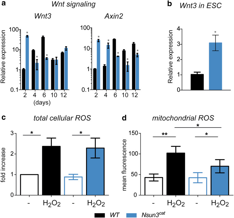
Nsun3 inactivation causes upregulation of Wnt signalling but does not affect basal ROS levels in ESCs. a RT-qPCR for Wnt3 and Axin2 was performed on cDNA prepared from EBs of wild-type and Nsun3 cat/cat cells at the indicated times of outgrowth on gelatine-coated plates. b Expression of Wnt3 is increased in Nsun3-mutant ESCs compared to wild-type cells. Normalization and quantification of PCR signals as in Fig. 6. c Quantification of total cellular ROS in control and H2O2-treated WT and Nsun3 cat/cat cells by FACS revealed no differences between the cell lines. Relative mean ± SEM values of 5 experiments are shown. d Quantification of mitochondrial ROS by MitoTracker Red CM-H2XROS staining and fluorescence microscopy showed weaker ROS induction after H2O2 treatment in Nsun3-mutant versus wild-type cells. Mean ± SEM intensity values of eight experiments are shown. (*p < 0.05; **p < 0.01)

