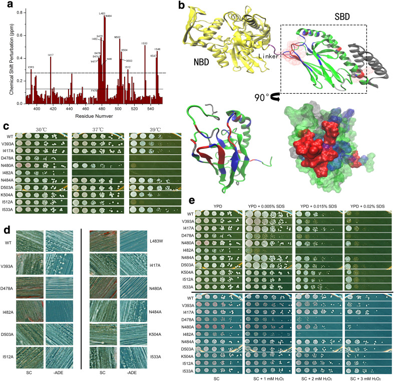
Fig. 5.
Interface regulates prion propagation and heat-shock response. a CSP histogram of L483W compared to WT at 30 °C. The solid line shows the average of the CSPs; the dotted line shows the average plus SD of CSPs. b Interface between SBD and NBD. Residues with significant CSPs were mapped onto the full-length DnaK structure (PDB:2KHO); overlay of SBDβ with residues in the SBD model of DnaK (PDB:1BPR) that have significant CSPs. Aspect was rotated by 90° as arrow indicates. The residues with significant CSPs over the average plus SD are in red; the residues with CSPs between the average plus SD and the average are in blue; the residues with CSPs below the average are in green; non-assigned residues are in grey. c Growth assay of predicted mutations at elevated temperatures. d Assessment of the predicted mutations on [PSI +] propagation. [psi −] cells were red colonies on YPD and unable to grow on −ADE plates; [PSI +] cells were white colonies on YPD and survived on −ADE plates. e Growth assay of predicted mutations under other stresses. YPD and SC medium were supplemented with cell-wall damage reagent SDS and oxidative damage reagent H2O2, respectively, to achieve required concentrations. Fresh cultures were spotted on those plates after a 1/5 serial dilution and incubated for 2 days at 30 °C

