
Figure 1.
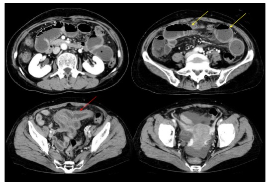
Abdominal computed tomography showing segmental edema of the intestinal wall (red arrow) with proximal dilatation (yellow arrow) and a small number of ascites.
Official websites use .gov
A
.gov website belongs to an official
government organization in the United States.
Secure .gov websites use HTTPS
A lock (
) or https:// means you've safely
connected to the .gov website. Share sensitive
information only on official, secure websites.
Abdominal computed tomography showing segmental edema of the intestinal wall (red arrow) with proximal dilatation (yellow arrow) and a small number of ascites.