Figure 1.

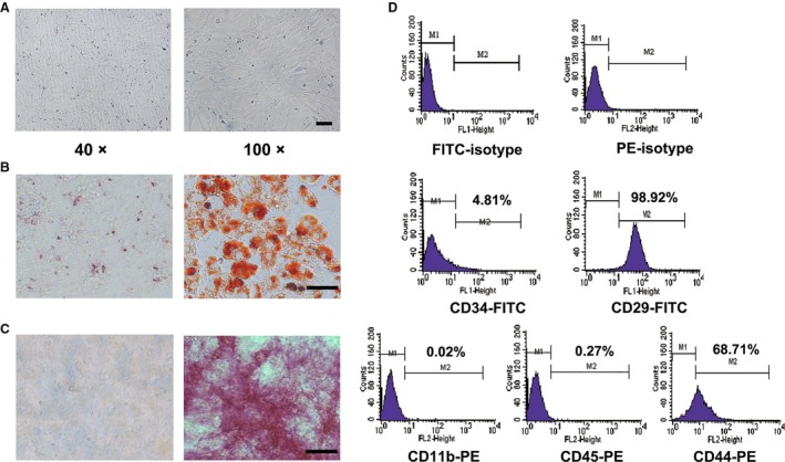
Morphology, phenotype, and multidifferentiation potentials of mMSCs. (A) Morphological appearance of murine bone marrow mesenchymal stem cells from 615 mice (mMSCs) at the third passage. Magnification: Left, ×40; Right, ×100, Scale bar = 100 μm. (B) Oil Red O staining detection for adipogenic differentiation. (C) Alkaline phosphatase staining detection for osteogenic differentiation. Magnification: ×200, Scale bar = 50 μm. (D) The surface antigens including CD34,CD29, CD11b, CD45, and CD44 of mMSCs detected by flow cytometry. Each surface markers’ positive rate was presented.
