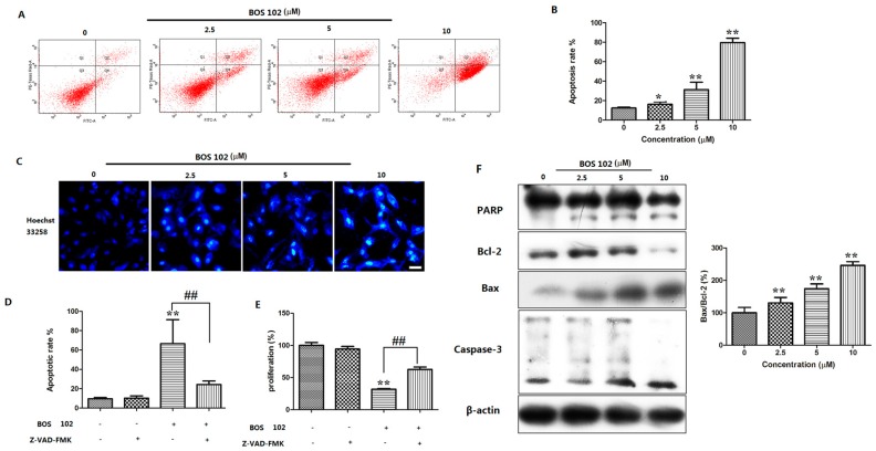
Figure 3.
BOS-102 induces intrinsic apoptosis in A549 cells. (A,B) FACS analysis via Annexin V/PI staining was used to identify apoptosis induced by BOS-102. A549 cells were treated with various concentrations of BOS-102 (0, 2.5, 5, 10 µM) for 48 h; (C) A549 cells were treated with BOS-102 (0, 2.5, 5, 10 µM) for 48 h. Hoechst 33258 staining was used to detected the apoptosis and photographed using fluorescence microscopy (Bar = 50 µm); (D) A549 cells were treated with 5 µM BOS-102 alone or in combination with Z-VAD-FMK (10 µM) for 48 h. The percentages of apoptotic cells were determined by flow cytometr (FACS) analysis via Annexin V/PI staining; (E) A549 cells were treated with 5 µM BOS-102 alone or in combination with Z-VAD-FMK (10 µM) for 48 h, cell viability was evaluated by MTT assay; and (F) Western blot analysis of apoptosis-related proteins, including PARP, Bcl-2, Bax, and Caspase-3. β-actin was used to normalize the protein content. The data represent mean values (±SD) obtained from three separate experiments. * p < 0.05, ** p < 0.01 vs. control group, ## p < 0.01 vs. 102(+)/Z-VAD-FMK(−) group.

