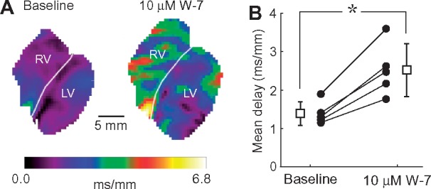
Figure 1.

Calmodulin blocker W7 depresses cardiac conduction in intact rabbit hearts. (A) Distribution of local conduction delay values in the imaged anterior surface of both ventricles at baseline (left) and after 30-min perfusion with 10 μM W7 (right). Local delays (the inverse of the local conduction velocities) were computed as the magnitude of the gradient of the activation time. Hearts were paced simultaneously from two electrodes located at the lateral epicardial surface of each ventricle at a cycle length of 300 ms. White labels indicate the LV and RV location, and the white lines indicate the boundary between the chambers. (B). Mean conduction delays computed over the entire mapped surface at baseline and after 30 min perfusion with 10 μM W7: solid circles represent values for individual experiments; open square symbols and bars represent average and SD. *P = 0.00352 vs. baseline, paired Student‘s t-test (n = 5 hearts).
