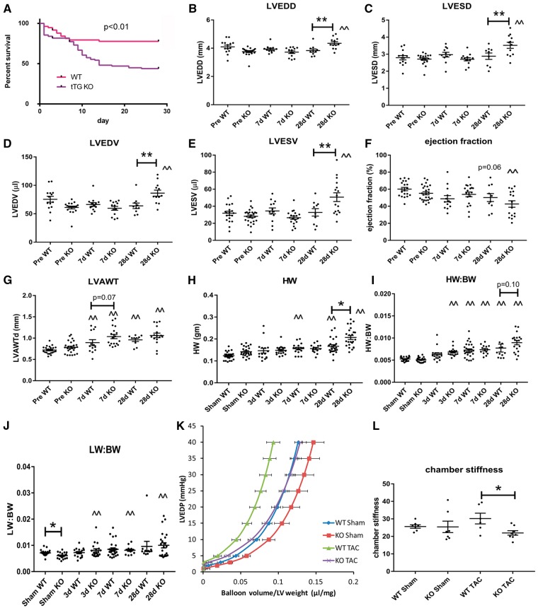
Figure 2.
tTG loss increases mortality and dilative ventricular remodelling, but protects from the development of diastolic dysfunction following pressure overload. (A) When compared with WT animals, tTG KO mice had increased mortality following pressure overload (P < 0.01; WT, n = 129; tTG KO, n = 128, log-rank test). (B–E) tTG KO mice had increased dilative remodelling after 28 days of TAC, exhibiting significantly higher LVEDD (B), LVESD (C), LVEDV (D) and LVESV (E) in comparison to WT animals (**P < 0.01 vs. corresponding WT, n = 13–18/group, one-way ANOVA followed by Sidak’s test). (F) tTG KO mice had a trend towards reduced ejection fraction after 28 days of TAC. (G) Both WT and tTG KO mice exhibited cardiac hypertrophy following pressure overload (^P < 0.05, ^^P < 0.01 vs. corresponding baseline values, Kruskal–Wallis test). tTG KO mice had trends towards increased anterior wall thickness after 7–28 days of TAC. (H and I) Both heart weight and heart weight:body weight ratio was increased in both WT and tTG KO animals following pressure overload (**P < 0.01 vs. corresponding sham, Kruskal–Wallis test). KO mice had significantly increased heart weight and a trend towards an increase in HW:BW after 28 days of TAC. (J) Lung weight:body weight ratio was also significantly increased in both WT and tTG KO mice after 7–28 days of TAC indicating the development of heart failure (*P < 0.05, **P < 0.01 vs. sham, n = 12–28/group, Kruskal–Wallis test). However, no significant differences in lung weight were noted between WT and tTG KO groups. (K and L) tTG loss attenuated diastolic dysfunction in the pressure overloaded myocardium. (K) Pressure:Volume analysis in isolated perfused hearts showed that tTG KO mice had a rightward shift of the curve. (L) Following TAC, chamber stiffness was significantly higher in WT animals when compared with tTG KOs (*P < 0.05, n = 6–8/group, ANOVA followed by Sidak’s test).

