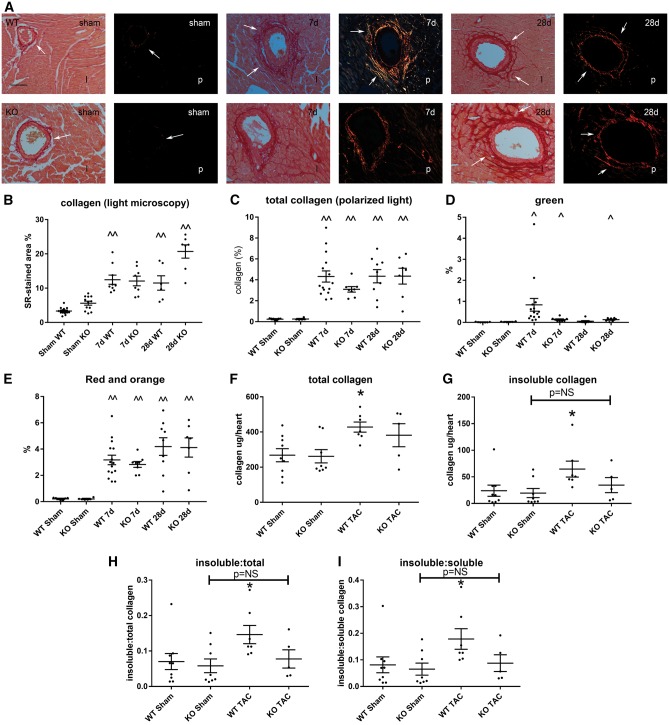
Figure 3.
Effects of tTG loss on collagen deposition in the pressure-overloaded myocardium. (A) Light microscopy (l) of sirius red-stained sections and polarized light microscopy (p) were used to study the distribution and birefringence of collagen fibres in sham and pressure-overloaded myocardium (arrows). (B and C) Quantitative analysis showed that in both WT and tTG KO animals, pressure overloaded hearts had significantly increased collagen-stained area (^P < 0.05, ^^P < 0.01 vs. corresponding sham, Kruskal–Wallis test, n = 6–14/group) after 7–28 days of TAC. No significant differences were noted in total collagen content (B and C) and in the amount of thinner green (D) and thicker orange-red (E) collagen fibres between WT and tTG KO groups. (F–I) A hydroxyproline biochemical assay showed that after 28 days of TAC, WT animals exhibited a significant increase in total myocardial collagen content (F), insoluble collagen (G), insoluble:total collagen (H) and insoluble:soluble collagen (I) when compared with sham mice (*P < 0.05 vs. sham, n = 6–9/group, Kruskal–Wallis test). In comparison to corresponding sham animals, tTG KO mice had no significant increase in total collagen content after 28 days of pressure overload and no significant increase in the fraction of insoluble collagen. The findings suggest that tTG loss may impair collagen cross-linking in the pressure-overloaded myocardium.

