Figure 2.
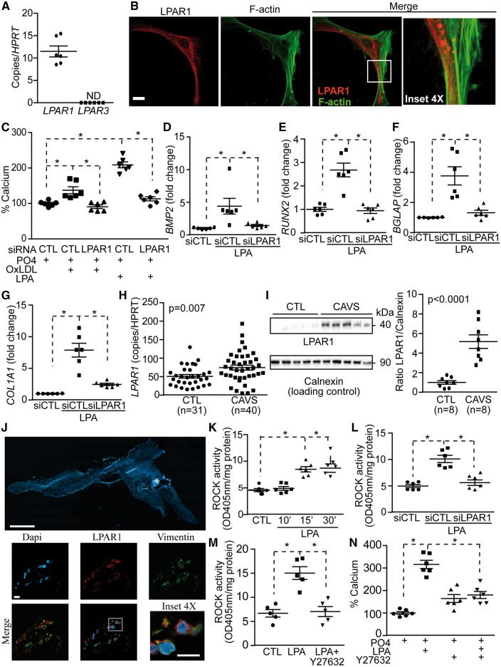
LPAR1 mediates LPA response in VICs. (A) LPAR1 expression in VICs (n = 6). (B) Confocal images of LPAR1 in VICs. Scale bar 20 µM, (n = 5). (C) LPAR1 is required for OxLDL and LPA induced mineralization of VICs (n = 6). (D–G) siLPAR1 prevented LPA-mediated rise of BMP2 (D), RUNX2 (E), BGLAP (F) and COL1A1 (G) (n = 6) (measurements at 24 h). (H) LPAR1 mRNA measurement in control non-mineralized (CTL) (n = 31) vs. calcified aortic valves (CAVS) (n = 40). (I) Representative western blot and quantification of LPAR1 in CTL (n = 8) vs. CAVS (n = 8). (J) Epifluorescence image of a calcified aortic valve showing the organization of the tissue in DAPI, scale bar 1000 µM and confocal images showing LPAR1 and vimentin co-expression in the same tissue, scale bar 10 µM (n = 10). (K–M) ROCK activity measurements; kinetics following LPA treatment (n = 6) (K), siLPAR1 negated LPA response (n = 6) (L) and Y27632 abrogated LPA effect (n = 5) (M). (N) Treatment with Y27632 reduces LPA-mediated mineralization in VICs (n = 6). Values are mean ± SEM. LPA: 10 µM, OxLDL: 100 ng/mL, Y27632: 5 µM; * P < 0.05.

