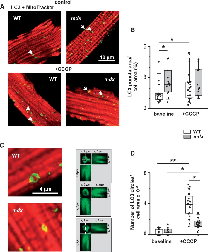
Figure 4.
Mitophagy is compromised in dystrophic cardiomyocytes isolated from hearts of 12+ months old mice (evidence from immunocytochemistry). (A) Representative overlay images of WT and mdx cardiomyocytes incubated with anti-LC3 antibodies and then with Alexa Fluor 488-conjugated secondary antibodies (green puncta on the image) and MitoTracker® Orange (red staining) without (top panels) and with (bottom panels) CCCP treatment. (B) Graph compares area occupied by LC3 puncta per cell area in myocytes with and without treatment with CCCP. Number of cells analysed was for WT (N = 3, n = 15 and N = 3, n = 24) and mdx hearts (N = 3, n = 15 and N = 3, n = 11) without and with incubation with CCCP, respectively. Data indicate enhanced autophagic activity in WT. Arrows on the images point to the ‘circle-like’ LC3 structures, which are likely to be associated with mitochondria. (C) Magnified images of cardiomyocytes containing the mitochondria associated LC3 circles. (D) Graph compares numbers of LC3 circles per cell area in WT (N = 3, n = 11 and N = 3, n = 21) and mdx (N = 3, n = 11 and N = 3, n = 20) myocytes with and without treatment with CCCP, respectively. Only LC3 puncta/circles associated with mitochondrial structures were considered for the analysis. This was ensured by taking additional images within ∼2 μm above and below the initial focal plane. Right panels in (C) show images of three representative mitochondria surrounded by LC3 puncta in three different projections (xy, xz, and yz). Data indicate that mitochondrial uncoupling induced substantially more mitochondria-associated LC3 circles in WT cardiomyocytes. Data obtained from mdx tissue are shown as gray bars whereas white bars illustrate data acquired from WT tissue. (*P < 0.05, **P < 0.01, ANOVA test).

