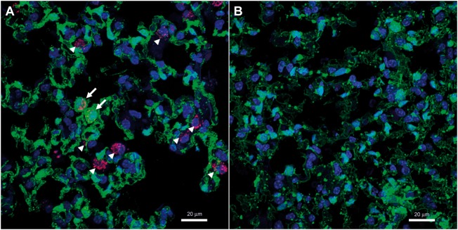
Figure 4.
Emergence of EPCs in the lung of Su/Hx-treated mice. (A) Confocal immunofluorescence image of c-kit+ cells (magenta red) in a Su/Hx mouse lung. Frozen sections of Su/Hx lungs were stained with a goat anti-mouse c-kit antibody and horse anti-goat DyLight 594. Arrows: ZsGreen+ c-kit+ cells; arrow heads: ZsGreen− c-kit+ cells.. (B) No c-kit+ cells were detected in a Nx lung following the same immunofluorescence staining and confocal imaging. Original magnification 800×; Magenta red, Immunostaining of c-kit; Green, fluorescence protein ZsGreen labelling of cells of endothelial origin; Blue, DAPI counterstaining of nuclei. The experiments were performed three times and the images shown are representative.

