Figure 1.
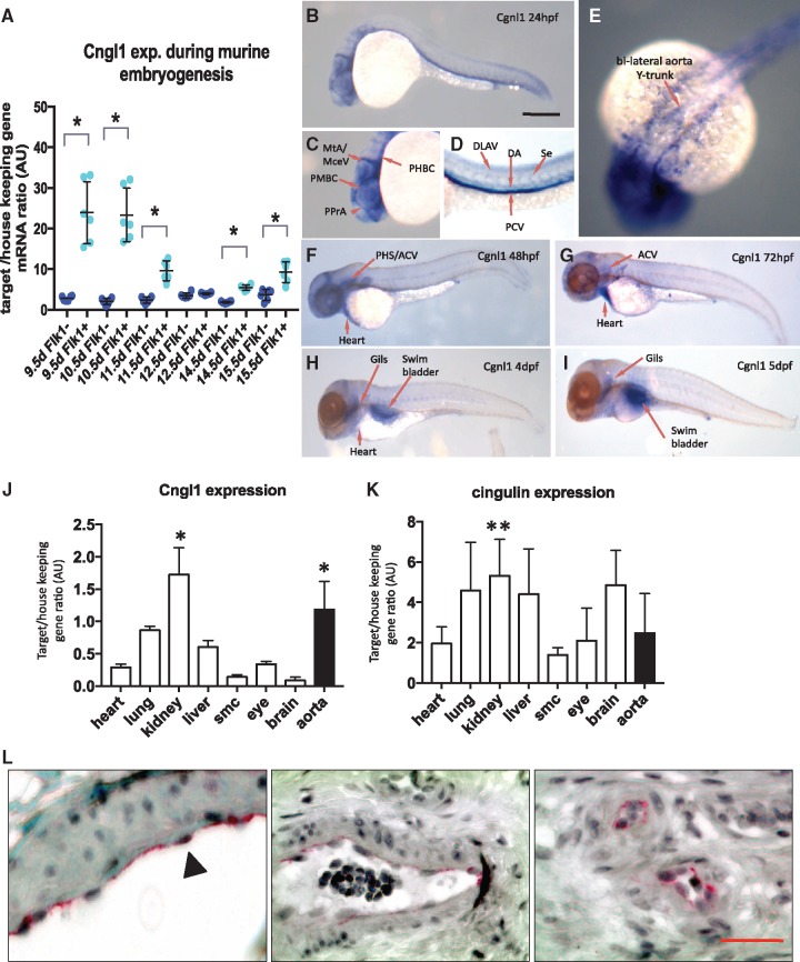
Cgnl1 is mainly expressed in vascular ECs. (A) Cgnl1 expression in Flk1+ and Flk1− cells during embryonic development of C57/bl6 mice from 9.5 to 15.5 days post-coital analyzed by qPCR. Mean ± SD, n = 6 for each time point, *P < 0.05 versus time point matched Flk1− cells. Student‘s t-test. (B) Whole-mount in situ hybridization of wild type zebrafish larvae at different time points. Scale bar represents 5 mm. Lateral view: Cgnl1 targeting antisense probe signal (blue) detected in the vasculature at 24 hpf. (C) Cgnl1 expression is visible in the cranial vessels, including the middle cerebral vein, metencephalic artery, primordial midbrain channel, primitive prosencephalic artery, and primordial hindbrain channel. (D) In the trunk region, Cgnl1 signal is in the posterior cardinal vein, dorsal longitudinal anastomotic vessel, DA, and intersegmental vessel (Se). (E) Dorsal view: Expression of Cgnl1 in the bilateral aorta Y-trunk. (F) Regression of Cgnl1 signal during larvae maturation: Lateral view: Cgnl1 expression at 48 hpf is mainly located in cranial vessels, primary head sinus and anterior cardinal vein (ACV), and heart region. (G) Cgnl1 expression at 72 hpf is still detected in the ACV, and remains visible in the heart, whereas expression of Cgnl1 at (H) 4 dpf and (I) 5 dpf is mainly in the gills and swimbladder. QPCR analysis of (J) Cgnl1 and (K) cingulin expression in various tissues of mature C57/bl6 mice. Mean ± SD, n = 5, *P < 0.05 aorta or kidney versus expression in all other tissues. **P < 0.05 kidney versus expression in skeletal muscle. One-way ANOVA, followed by post hoc test with aorta or kidney set as control. (L) Immunostaining of Cgnl1 in human paraffin sections: From left to right, Cgnl1 expression is observed in the endothelium of macrovessels (first and second micrograph, indicated by black arrowhead) and microvessels (third micrograph). Scale bar represents 50 µm. Red signal, VEGFR2 immuno-staining; black, nucleus staining.

