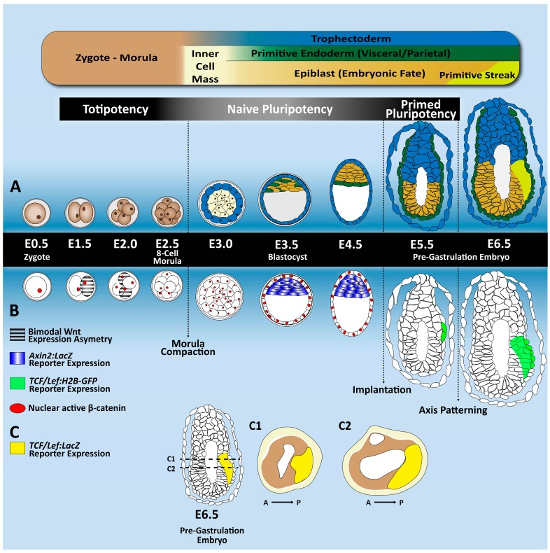
Figure 2.
Tracing Wnt activity across early mouse embryogenesis. (A) A schematic overview of the mouse pre-implantation and post-implantation developmental stages, from zygote formation (E0.5) until the pre-gastrulation stage (E6.5). After fertilization, the zygote undergoes a series of mitotic divisions together with progressive cell fate acquisition. At the end of the morula stage, the first segregation event occurs giving rise to the trophectoderm and the inner cell mass (ICM). At E4.5–E5.0, after the ICM segregates into the epiblast and primitive endoderm, the blastocyst implants in the uterus. Around E6.5, the egg cylinder is formed, and anterior–posterior axis patterning is established, along with the first mesendodermal progenitors at the primitive streak. (B) This chart provides information about the main molecular changes in the Wnt/β-catenin signaling pathway. The bimodal Wnt target gene expression is represented in grey stripes and is (at transcript level) already detected at the two- and four-cell stages [27]. In red, active nuclear β-catenin expression [28]. In blue, the Axin2:LacZ reporter is found only at the blastocyst stage [29]. In green, detection of the TCF/Lef:Histone 2B-green fluorescent protein (H2B-GFP) reporter occurs only after implantation stages [30]. (C) Longitudinal and transversal sections of a pre-gastrulating mouse embryo (E6.5) showing in yellow the distribution of the TCF/Lef:β-galactosidase reporter activity in the posterior region [30].

