Figure 4.
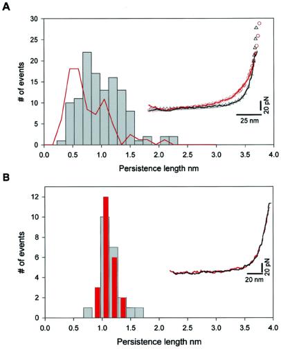
The PEVK segment of cardiac titin shows multiple mechanical conformations. (A) Frequency histogram of the measured persistence length of the PEVK segment (gray bars). The force-extension relationships are accurately described by the WLC model of polymer elasticity (as shown in the Inset). The close agreement between the WLC model and the data (within ≈5 pN) demonstrates that extension of the PEVK segment does not involve the rupture of hydrogen-bonded structures. The PEVK segment shows a wide range of persistence length values, in agreement with the persistence lengths calculated from the end-to-end distributions observed with EM (red line). (Inset) Two representative PEVK recordings with different persistence lengths. In these two traces only the initial length of a stretched (I27-PEVK)3 polyprotein (before I27 domain unfolding) is shown (solid lines are experimental recordings, open symbols are Levenberg–Marquardt nonlinear fit of WLC model to the individual recordings). The red recording has a persistence length of 0.40 nm, the black recording has a persistence length of 1.08 nm. For comparison, the red recording (with a contour length of 207 nm) was normalized to have the same contour length as the black trace (140 nm). (B) Frequency histogram of the measured persistence length of the PEVK segments of a single (I27-PEVK)3 molecule during repeated stretch and relaxation cycles. The persistence length is narrowly distributed around 1.1 nm and is the same for the stretch (gray bars) and the relaxation (red bars) traces, showing that there is no detectable change in persistence length during these cycles. The scatter is due to the error margin of the fits to the data. (Inset) Two consecutive stretch (black) and relaxation (red) recordings. No hysteresis between stretching and relaxation was observed, indicating that this process is fully reversible.

