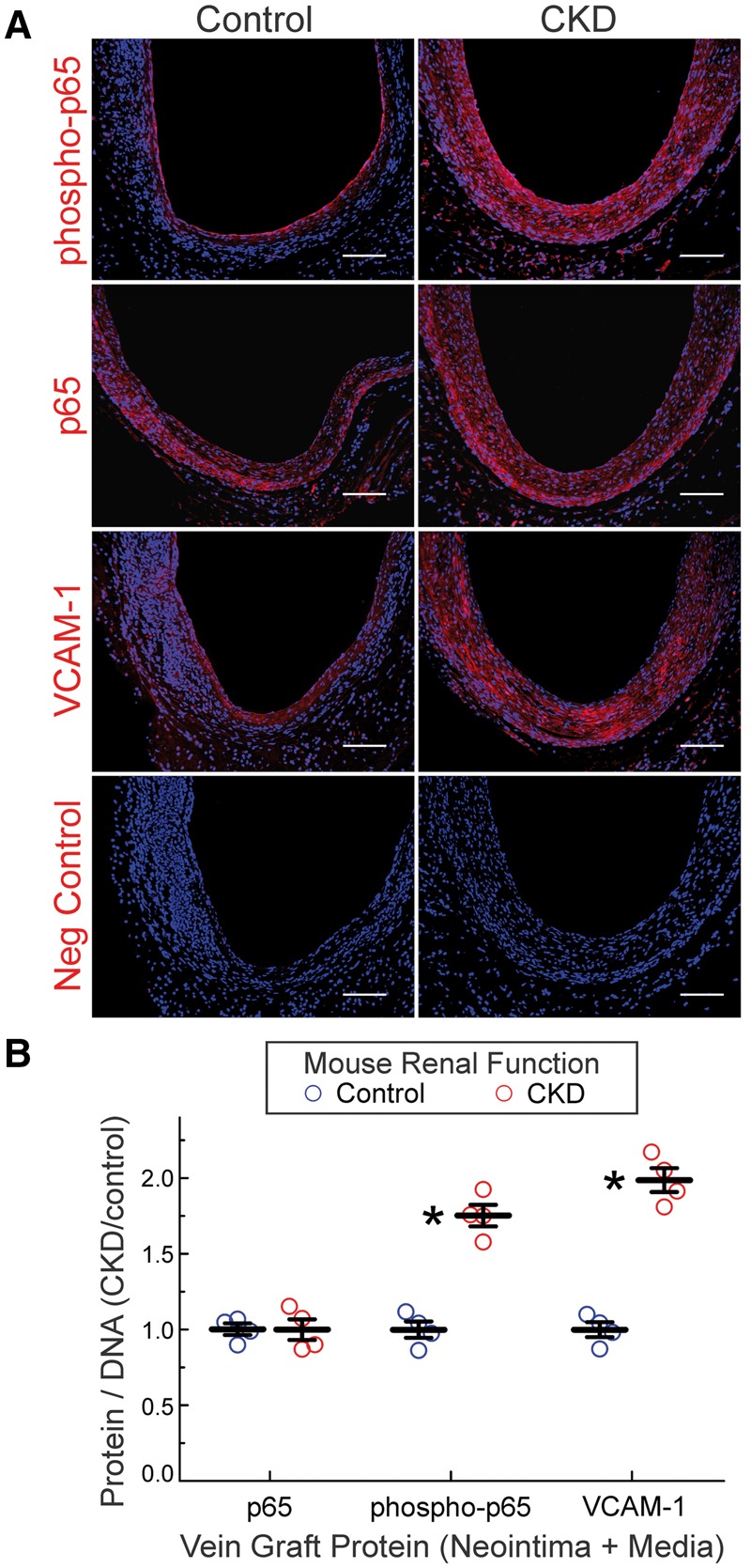
Figure 2.

CKD augments NFκB activation in vein grafts. (A), Serial sections of vein grafts from Figure 1 were immunostained with rabbit IgG targeting the following proteins: NFκB p65 subunit phosphorylated on Ser536 (phospho-p65); NFκB p65 subunit, phosphorylated or not (p65); VCAM-1; or no specific protein (Neg Control). Alexa 546-conjugated anti-rabbit IgG and Hoechst 33 342 were used on all specimens. Specimens from control and CKD mice were stained concurrently. (B), The ratios of red (protein) to blue (DNA) pixels in the neointima plus media were quantitated by Image J, and normalized to the cognate ratios obtained for control specimens in each staining cohort, to obtain ‘CKD/control’, plotted individually and as the means ± SE for each group. Compared with control: *P < 0.01 (2-way ANOVA with post-hoc Sidak test).
