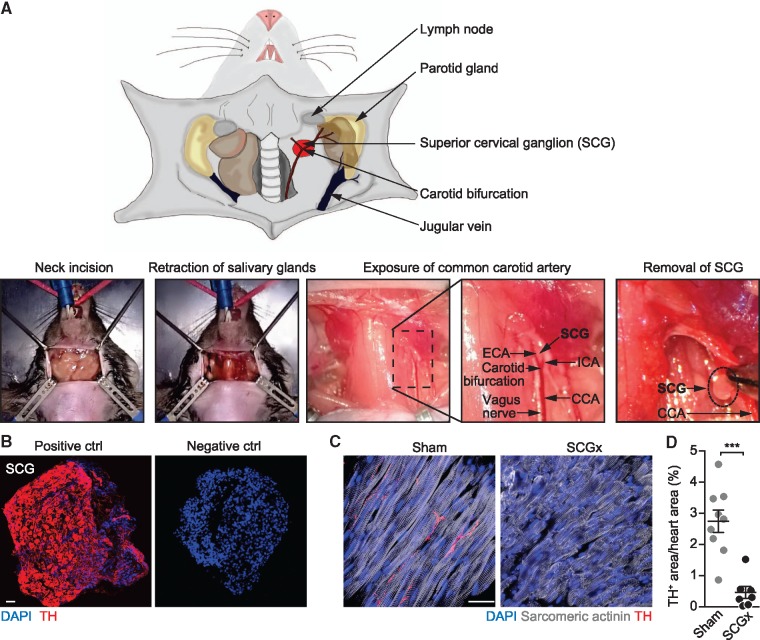
Figure 1.
Surgical removal of the SCGs in mice. (A) (Upper panel) Scheme depicting the anatomical location of the SCG (red) and neighbouring structures. For better overview, the sternocleidomastoid muscle has not been pictured. (Lower panels) Surgical procedure of SCG removal (SCGx). A vertical incision is applied to the skin of the clean-shaved neck at the height of the cervical vertebrae C1/C2. Exposed glandular tissue and the sternocleidomastoid muscle are then pulled to the side to expose the CCA. The SCG is located behind the bifurcation of the CCA into the internal (ICA) and the external carotid artery (ECA). Next to the CCA, the vagal nerve is situated. Finally, the preganglionic and postganglionic nerves of the SCG are cut with a scissor and the ganglion is removed. (B) Immunofluorescent staining of surgically removed SCG. The same staining procedure served as a negative control, except that the primary antibody was omitted. Tyrosine hydroxylase (TH), scale bar: 50 μm. (C) Heart sections stained for sympathetic nerves (TH) in mice with ganglionectomy (SCGx) and control mice (sham), scale bar: 20 μm. (D) Quantification of TH-positive area. Data are mean ± SEM of n = 9 (sham) and n = 7 mice (SCGx). Student’s t-test was applied for statistical analysis. ***P < 0.001.

