Figure 2.
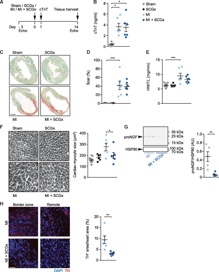
Sympathetic denervation attenuates cardiac hypertrophy after MI. (A) Experimental set-up. Intervention groups included sham, SCGx (bilateral removal of the SCG), MI (myocardial infarction by LAD ligation), and MI + SCGx (bilateral SCG-removal followed by LAD ligation). Three days prior to surgical intervention, cardiac function was assessed by echocardiography. Twenty-four hours after surgery, plasma troponin levels were measured. Functional analysis and tissue harvesting was performed 14 days after the intervention. (B) Cardiac troponin T (cTnT) values determined in plasma 24 h after surgery. (C) Myocardial paraffin sections stained with Fast Green/Sirius Red. Scale bar: 1 mm. (D) Quantification of Sirius Red-positive area. (E) Heart weight-to-tibia length ratio. (F) WGA staining for quantification of cardiac myocyte dimensions. Scale bar: 50 μm. (G) Representative immunoblot of pro-NGF expression in LV myocardium after MI and MI + SCGx and quantitative analysis of the data. (H) Quantification of intramyocardial nerves within the LV anterior wall by staining with an antibody directed against TH. Data are mean ± SEM of n = 9 (sham), n = 7 (SCGx) and n = 6 mice (MI, MI + SCGx). cTnT values were analysed with the Kruskal–Wallis-test. *P < 0.05 vs. respective sham. Two-way analysis of variance with Bonferroni’s multiple comparison test was applied for statistical analysis of scar formation, heart weight-to-tibia length ratio and cardiac myocyte size. *P < 0.05, **P < 0.01, ***P < 0.001. Mann–Whitney test was used for analysis of pro-NGF/HSP90 and TH-positive area. ***P < 0.001 vs. MI.

