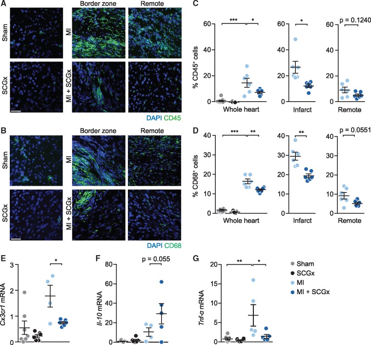
Figure 3.
Reduced inflammatory cell infiltration upon sympathetic denervation. (A, B) Representative confocal images of LV tissue sections 14 days after the respective intervention stained with DAPI and anti-CD45 antibody (A) or anti-CD68 antibody (B). (C) Quantification of CD45- and (D) CD68- cells (% of DAPI-positive cells) in whole heart, infarct and remote area. Scale bar: 50 μm. Data are mean ± SEM of n = 9 (sham), n = 7 (SCGx), and n = 6 mice (MI, MI + SCGx). (E–G) Determination of inflammation-related mRNAs by PCR in total RNA prepared from LV myocardium from the indicated groups. Relative expression of (E) CX3C chemokine receptor 1 (Cx3cr1), (F) interleukin-10 (Il-10) and (G) tumour necrosis factor alpha (Tnf-α). The mRNA levels were first normailzed to Hprt and then further normalized to the levels in sham. All four groups were compared by two-way analysis of variance with Bonferroni’s multiple comparison test. *P < 0.05, **P < 0.01, ***P < 0.001. MI and MI + SCGx groups were analysed with Student’s t-test. *P < 0.05 and **P < 0.01 vs. MI.

