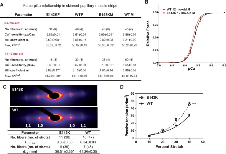
Figure 4.
E143K-induced changes of function and sarcomere lattice in papillary muscle strips. (A) Development of steady-state force in skinned papillary muscle fibers from E143K vs. WT mice. Two age groups with 3–6 animals per group were used. (B) Representative force-pCa relationship in ∼12 mo-old E143K-M vs. WT-M mice. (C) Fiber diffraction pattern in ∼6 mo-old male E143K (4M, 2F) vs. WT (5M, 1F) mice under relaxation conditions (pCa 8). Lattice spacing d1,0 (in nm) and equatorial reflections’ intensity ratio I1,1/I1,0. Sarcomere length was 2.1 µm. Note a significant decrease in the lattice spacing in the mutant mice compared with WT mice. Data are expressed as average ± SEM of n (number of fibers) with *P < 0.05 vs. WT (t-test). (D) Passive tension measured in ∼6 mo-old E143K (2M, 1F yielding 29 fibers) vs. WT (2F, 1M yielding 27 fibers). Note significantly higher passive tension measured in E143K vs. WT hearts as assessed by two-way ANOVA for repetitive measurements (**P < 0.01).

