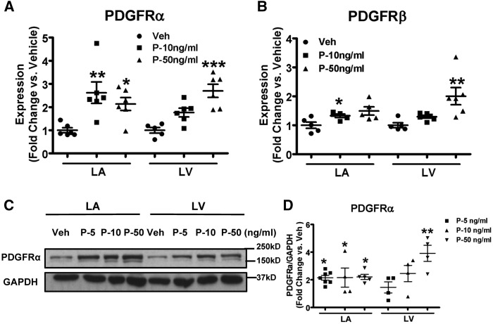
Figure 3.
Exposure to PDGF upregulates PDGFRs in canine fibroblasts. (A,B) mRNA expression of PDGFRα and PDGFRβ in atrial and ventricular fibroblasts stimulated with PDGF-AB at different concentrations. (C) Representative immunoblots for PDGFRα in LA and LV fibroblasts stimulated with PDGF-AB. (D) Band-intensities for PDGFRα normalized to GAPDH and presented as fold-change versus corresponding vehicle. Mean ± S.E.M., n = 4–7/group for A, B, and D. All values were normalized to corresponding vehicle on the same blot for D. *P < 0.05, **P < 0.01, ***P < 0.001 vs. corresponding vehicles. One-way ANOVA with Dunnett’s tests were used for statistical analysis. LA, left atrial; LV, left ventricular; FB, fibroblast; Veh: vehicle; P, PDGF.

